ULTRASOUND SERVICES
What Is Veterinary Diagnostic Ultrasound?
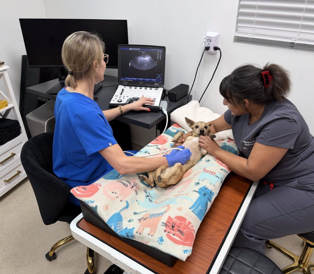
Ultrasound is a safe, painless, and non-invasive imaging technique that allows specialist veterinarians to see inside your pet’s body in real time. It uses sound waves—rather than radiation—to create detailed images of organs, tissues, and blood flow. Ultrasound is commonly used to evaluate the abdomen, chest, heart, and other soft tissues in dogs and cats.
How Does Ultrasound Work?
During an ultrasound exam, a small handheld probe is gently placed on your pet’s skin. The probe sends out high-frequency sound waves that bounce off internal structures such as the liver, kidneys, intestines, bladder, or spleen. These returning sound waves are converted into live images on a screen, allowing the radiologist to see how organs look and function in real time. Ultrasound is more accurate than radiographs for diagnosing obstructions in the gastrointestinal tract as well as issues with solid organs (e.g. liver). Veterinarians may use x-rays as a first line diagnostic and then follow up with a complete ultrasound to obtain more information. Because ultrasound shows motion—such as blood flow or organ movement—it can also provide additional information that other imaging methods cannot.

How Ultrasound Helps with Accurate Diagnosis?
Ultrasound is especially valuable for diagnosing conditions that affect soft tissues. It can help detect abnormalities such as stones, tumors, obstructions, cysts, inflammation, fluid buildup, organ enlargement, intestinal disease, and changes in blood flow. In many cases, ultrasound also allows for guided sampling (such as aspirates or biopsies), improving diagnostic accuracy while minimizing discomfort for your pet. By providing detailed, real-time information, ultrasound helps veterinarians make faster, more precise diagnoses and develop the most appropriate treatment plans.
Reasons to Perform an Ultrasound
Your veterinarian may refer you and your pet to Eclipse Veterinary Imaging for an ultrasound in order to determine the cause of your pet’s symptoms. These may include vomiting, diarrhea, weight loss, lethargy/ low energy, hiding, jaundice, as well as to determine the cause of abnormalities found on blood work (e.g. elevated liver enzymes or kidney values).
Expertise You Can Trust
All ultrasound examinations at Eclipse Veterinary Imaging are performed and interpreted by board-certified veterinary radiologists, certified by the American College of Veterinary Radiology (ACVR). This advanced level of training ensures that every study is conducted with the highest standards of accuracy, consistency, and medical expertise—giving your veterinarian clear, reliable answers and giving you confidence in your pet’s care.
What does “board-certified” mean?
“An American College of Veterinary Radiology (ACVR) board-certified veterinary radiologist is an individual who has undergone advanced training, completed the requirements, and passed the ACVR certification examination (written and oral) to become certified as a specialist in veterinary radiology (an ACVR Diplomate). A number of certifying organizations for veterinary medicine are recognized by the American Veterinary Medical Association (AVMA). It is possible that a doctor is board-certified, but in specialty other than veterinary radiology (diagnostic imaging). These specialties might include internal medicine or surgery, for example. Although these individuals are board-certified by organizations recognized by the AVMA, they do not have the advanced training and expertise required for veterinary imaging and interpretation (radiology, ultrasound, computed tomography (CT), magnetic resonance imaging (MRI), nuclear medicine) or radiation therapy. Individuals from other groups who call themselves ‘Diplomates’ or who claim ‘Certification’ may not be from organizations recognized by the AVMA.” From https://acvr.org/faqs/